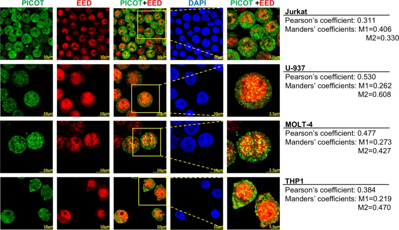
Fig. 1. Nuclear PICOT partially colocalizes with EED in leukemia cells.
Jurkat, U-937, MOLT4 and THP1 cells (1.5 × 10⁶/group) were seeded on poly-L-lysine-coated 8 well µ-slide (ibidi Ltd.) and deposited by centrifugation at 1200 rpm for 5 min. The cells were fixed, permeabilized and incubated with a mixture of mouse anti-PICOT mAbs and rabbit anti-EED polyclonal Abs for 1 h at room temperature. The cells were then stained using Cy3-conjugated anti-mouse Abs and Cy5-conjugated anti-rabbit Abs, plus nuclei counterstain with DAPI (1 µg/ml in PBS) for 1 h in the dark, at room temperature. The cells were then analyzed using a confocal microscope (Olympus Flouview 1000 laser scanning confocal microscope). PICOT-EED colocalization is demonstrated in a color overlay panel, and a selected area marked by a red box that was magnified is shown in the right panel. Comparative quantification of PICOT-EED colocalization was performed using the ImageJ colocalization JACoP imageJ plugin18. Pearson’s coefficient value and Manders’ coefficient values (M1 = red overlap with green; M2 = green overlap with red) are indicated on right for each panel

