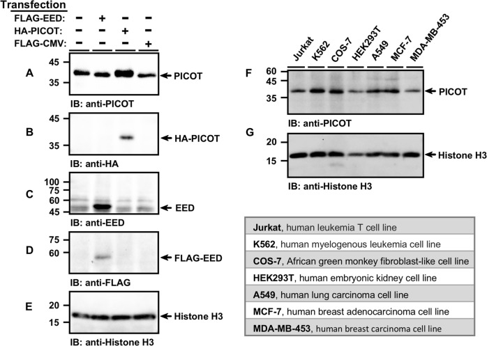
Fig. 2. PICOT reside in the chromatin fraction of tumor cell lines.
COS-7 cells were transiently transfected with the indicated expression vectors using the PEI reagent. Chromatin lysates from transfected and untransfected COS-7 cells were prepared using the protein-protein ChIP protocol, boiled, and subjected to SDS-PAGE (5 µg/lane) on two parallel 12.5% gels under reducing conditions. Proteins were then electroblotted onto two parallel nitrocellulose membranes that were immunoblotted with either mouse anti-PICOT mAbs (a), or rabbit anti-HA polyclonal Abs (b), followed by development using the immunoperoxidase ECL detection system and autoradiography. The membranes were then immunoblotted with rabbit anti-EED Abs (c), mouse anti-FLAG mAbs (d) and mouse anti-Histone H3 Abs (e), which served as a protein loading control. In a similar experiment, chromatin lysates were prepared from seven different cell lines and samples were immunoblotted with mouse anti-PICOT mAbs (f) and mouse anti-Histone H3 mAbs (g). The origin of the cell lines is indicated in the table below

