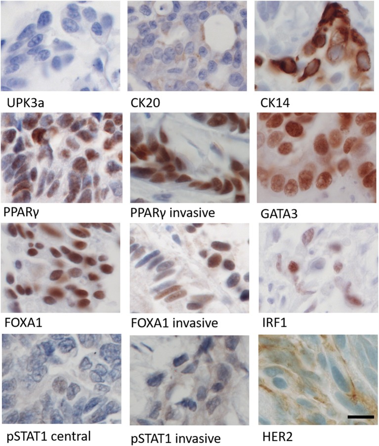
Figure 5.
Immunohistochemical labeling of patient's tumor using panel of antibodies against differentiation (UPK3a, CK20, CK14), transcription factor (PPARγ, GATA3, FOXA1, IRF1), and cell signaling (pSTAT1 and HER2) markers. Images from central region of the tumor, except where marked as invasive. Parallel human tissue controls for antibodies are shown in Figure S3. Scale bar = 12 μm.

