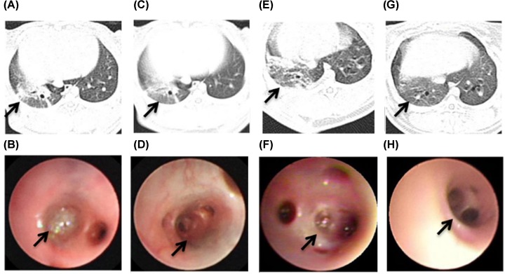
Figure 6. The performance of CT and bronchoscopy after co-therapy of hUMSCs combined with LZD in the MRSA-infection rabbit pneumonia.
The area of pulmonary consolidation of LZD group (arrow) was observed by CT at 48 (A) and 168 h (C). The purulent secretion congestion (arrow) could be seen in the bronchus of right lower lobe under the bronchoscope at LZD group at 48 (B) and 168 h (D). The pulmonary consolidation (arrow) of combined group was observed by CT at 48 (E) and 168 h (G). The mucosal hyperemia and erosion (arrow) of combined group of LZD group at 48 (F) and 168 h (H) was observed under the bronchoscope.

