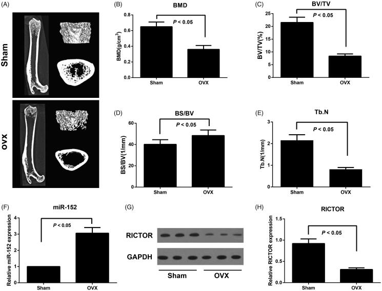
Figure 1.
Bone microarchitectural changes in femur assessed by micro-CT and expression of miR-152 and RICTOR in ovariectomized rat model of osteoporosis. Note: A, Representative micro-CT images of the right femur; B, Bone mineral density (BMD) of femoral tissues was measured; C–E, Micro-CT analysis of BV/TV (C), BS/BV (D), and Tb.N (E) in both OVX and Sham rats; BV/TV, Bone volume/total volume; BS/BV, Bone surface/bone volume; Tb.N, trabecular number; F, The miR-152 levels in femoral tissues in both OVX and Sham rats detected by qRT-PCR; G-H, RICTOR protein expression in femoral tissues in both OVX and Sham rats detected by Western blot.

