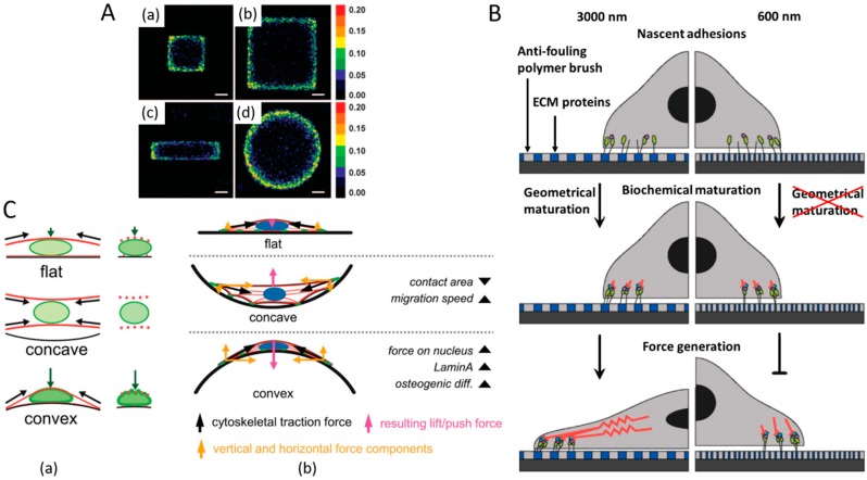
Figure 2.
The effects of substrate geometry on cells. (A) The patterns of forces exerted by the cells responding to the edges and boundaries of different substrates. (a) Colorimetric stacked images of cell proliferation in a small (250 µm edge) square, (b) large (500 µm edge) square, (c) small (125 × 500 µm) rectangular, and (d) large (564 µm diameter) circular islands [37]. “Reprinted with permission from ref. [37]. Copyright 2005, National Academy of Sciences” (B) A model of geometrical, biochemical, and mechanical maturation of integrin-mediated cell adhesion and behaviour after responding to nanopatterned matrices [42]. “Reprinted with permission from [42]. Copyright 2014, American Chemical Society.” (C) Schematic representation of (a) the cytoskeletal forces acting on the nucleus (F-actin in red and lamin-A in green) and (b) the proposed geometry-induced changes in cellular attachment and forces on the nucleus for flat, concave and convex surfaces [43]. “Reprinted with permission from ref. [43]. Reproduced with permission under Creative Commons Attribution 4.0 International License http://creativecommons.org/licenses/by/4.0/.

