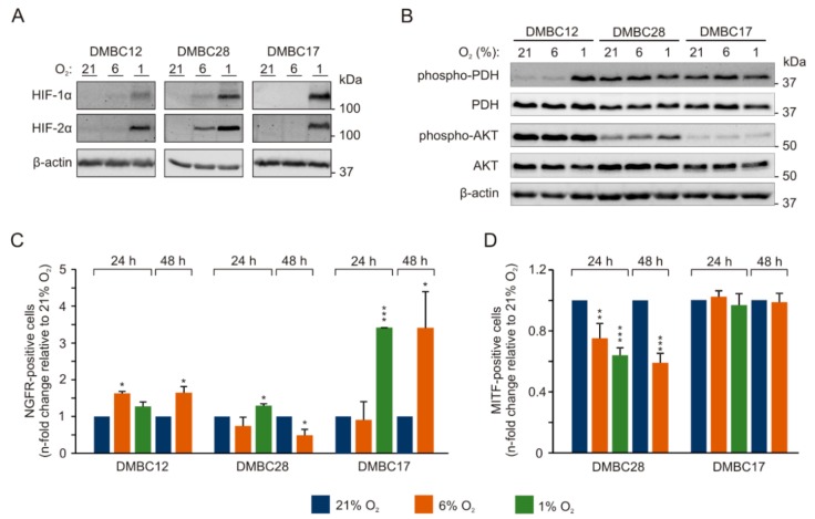
Figure 2.
Phenotypic alterations in melanoma cells in different oxygen concentrations depend on initial phenotype of melanoma cells. Melanoma cells were incubated at different oxygen concentrations, 21% O2 (hyperoxia) 6% O2 (normoxia) and 1% O2 (hypoxia). Protein levels of (A) HIF (hypoxia-inducible factor)-1α and HIF-2α and (B) p-PDH (phosphorylated pyruvate dehydrogenase, inactive form), PDH (total), p-AKT (phosphorylated protein kinase B) and total AKT were assessed by immunoblotting with representative images shown. β-actin was used as a loading control. The percentages of (C) NGFR (nerve growth factor receptor)-positive cells and (D) MITF (microphthalmia-associated transcription factor)-positive cells in normoxia and hypoxia relative to the standard culture conditions (21% O2). MITF-positive cells were almost undetectable in DMBC12 cell population (see Figure S1). n = 3, except for hypoxia (n = 2). Differences are considered significant at * p < 0.05, ** p < 0.01, *** p < 0.001.

