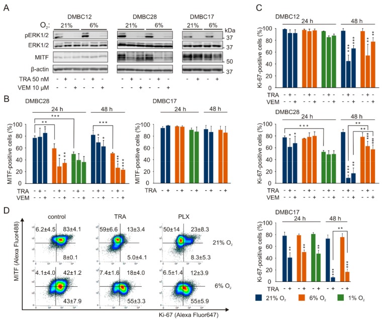
Figure 5.
Vemurafenib and trametinib while targeting the MAPK/ERK pathway irrespective of oxygen concentration, affect MITF-positive and Ki-67-positive melanoma cells in oxygen- and cell line-dependent manner. (A) The effects of trametinib (TRA) and vemurafenib (VEM) on ERK1/2 activity and MITF level in 21% and 6% O2 were determined by Western blotting with representative images shown (n = 3). β-actin was used as a loading control. (B) The percentage of MITF-positive cells in control cultures and after treatment with either 50 nM trametinib (TRA) or 10 µM vemurafenib (VEM) are shown as mean values ± SD (n = 3). Results for DMBC12 were not quantified due to almost undetectable MITF level in control and lack of changes in drug-treated cells. Differences are considered significant at * p < 0.05 ** p < 0.01 *** p < 0.001. See Figure S1 for representative dot plots. (C) The percentage of Ki-67-positive cells in control cultures and after treatment with 50 nM trametinib or 10 µM vemurafenib are shown as mean values of 3 biological replicates ± SD. Differences are considered significant at * p < 0.05 ** p < 0.01 *** p < 0.001. See Figure S1 for representative dot plots. (D) Representative density plots of dual-stained DMBC28 cells with anti-MITF and anti-Ki-67 antibodies after 48 h of drug treatment. Average percentages of cells positive for one or both markers are shown (n = 2).

