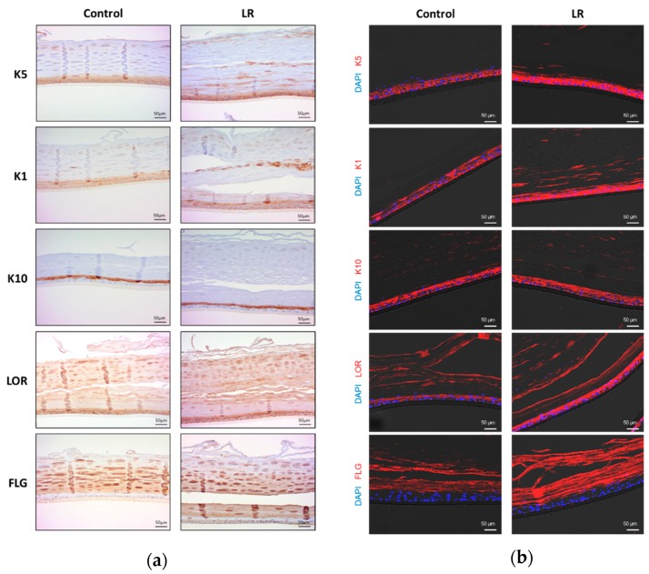
Figure 3.
Immunohistochemical and immunofluorescence images of KeraskinTM treated with LR lysate. (a) Immunohistochemical images of KeraskinTM with antibodies against cytokeratin 5 (K5), 1 (K1), 10 (K10), loricrin (LOR) and filaggrin (FLG). LR lysate was topically applied to KeraskinTM every other day for 16 days. (b) Immunofluorescence images of KeraskinTM. DAPI (blue) was used for nuclear staining and red fluorescent indicated lineage markers.

