Figure 1.
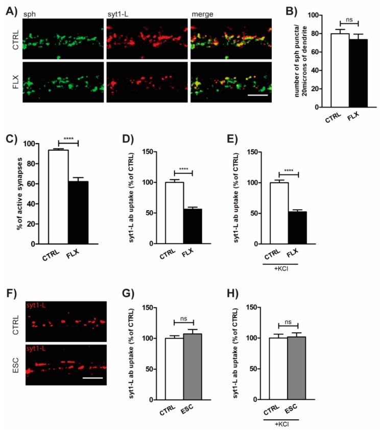
Fluoxetine (FLX) reduces presynaptic activity. (A) Representative images of primary cortical neurons treated with FLX (100 µM: 90 min) or control solution (CTRL) and stained with antibody against presynaptic marker synaptophysin (sph; green), syt1-L ab (red), and merged. (B) Quantification of the number of sph-positive puncta along 20 µm of proximal dendrite in CTRL (n = 29) and FLX-treated cells (n = 31). (C) Quantification of the percentage of active synapses in CTRL (n = 29) and FLX-treated neurons (n = 31). Active synapses were determined as puncta positive for both sph and syt1-L ab along 20 µm of proximal dendrite. (D) Quantification of “spontaneous” (network-activity-driven) syt1-L ab uptake along 20 µm of proximal dendrite (n = 37 CTRL cells; n = 39 FLX-treated cells). (E) KCl-evoked syt1-L ab uptake along 20 µm of proximal dendrite in CTRL (n = 41) and FLX-treated cells (n = 41). (F) Representative images and (G) corresponding quantification of syt1-L ab uptake under “spontaneous” condition (n = 35 CTRL cells; n = 34 ESC-treated cells) or (H) KCL-evoked condition upon the treatment of cells with 100 µM ESC (n = 25 cells) or CTRL solution (n = 28 cells). In all graphs, bars denote intensity values ± standard error of the mean (SEM) normalized to the mean intensity value in the control group. The statistic was assessed using Mann–Whitney test (B,C,G,H) or Student’s t-test (D,E,G); **** p < 0.0001. Scale bar, 5 µm.

