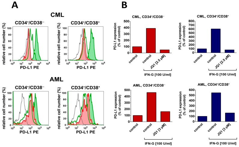
Figure 2.
Expression of PD-L1 on leukemic stem cells and regulation by the BRD4/MYC blocker JQ1.A: Mononuclear cells (MNC) were obtained from the bone marrow of a patient with chronic leukemia (CML, upper panel) and from the blood of a patient with acute myeloid leukemia (AML FAB M4, lower panel). Both patients gave written informed consent before BM aspiration was performed. The study was approved by the ethics committee of the Medical University of Vienna. MNC were incubated in control medium (open green histogram), recombinant IFN-G (100 U/mL; green histogram) or in a combination of interferon γ (IFN-G) (100 U/mL) and 3 μM of JQ1 (AML) or IFN-G (100 U/mL) and 2.5 µM of JQ1 (CML) (red histograms) for 24 h at 37 °C. Then, the expression of PD-L1 on CD45+/CD34+/CD38─ LSC (left panels) and CD45+/CD34+/CD38+ stem and progenitor cells (right panels) was measured by a monoclonal antibody against PD-L1 on a FACSCalibur (BD Biosciences). The isotype-matched control antibody is shown as a grey histogram. B: shows the IFN-G-induced upregulation of PD-L1 (compared to medium control = control) on LSC (red bars, left panels) and on CD34+/CD38+ leukemic progenitors (blue bars, right panels) as a bar diagram in one patient with chronic phase CML (upper panels) and one patient with AML (FAB M4, lower panels) and the effects of JQ1 (3 µM) on IFN-G-induced upregulation of PD-L1 in these cells. FAB, French–American–British cooperation study group classification.

