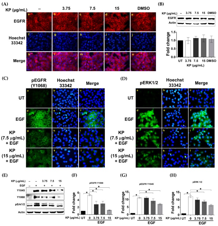
Figure 4.
(A) The effects of KP on EGF receptor (EGFR) expression in HeLa cells treated with different concentrations of KP or DMSO for 24 h; (B) Western blotting with quantitative analysis of total EGFR in HeLa cells treated with different concentrations of KP or DMSO for 24 h; (C) immunofluorescence for EGFR phosphorylation (pEGFR) at tyrosine 1068 (Y1068) (green) in HeLa cells treated with different concentrations of KP for 24 h and stimulated with EGF for 2 min; (D) immunofluorescence for ERK1/2 phosphorylation (green) in HeLa cells treated with different concentrations of KP for 24 h and stimulated with EGF for 15 min. Nuclei were counterstained with Hoechst 33342 (blue); (E) Western blot analysis for EGFR phosphorylation at Y1045 and Y1068 and pERK1/2 in HeLa cells treated with different concentrations of KP for 24 h and stimulated with EGF for 2 min; (F) quantitative analysis for EGFR phosphorylation at Y1068; (G) quantitative analysis for EGFR phosphorylation at 1045; (H) quantitative analysis for ERK1/2 phosphorylation. Micrographs were captured at 40× magnification. Data represent mean ± SD of three replicates. * p < 0.05.

