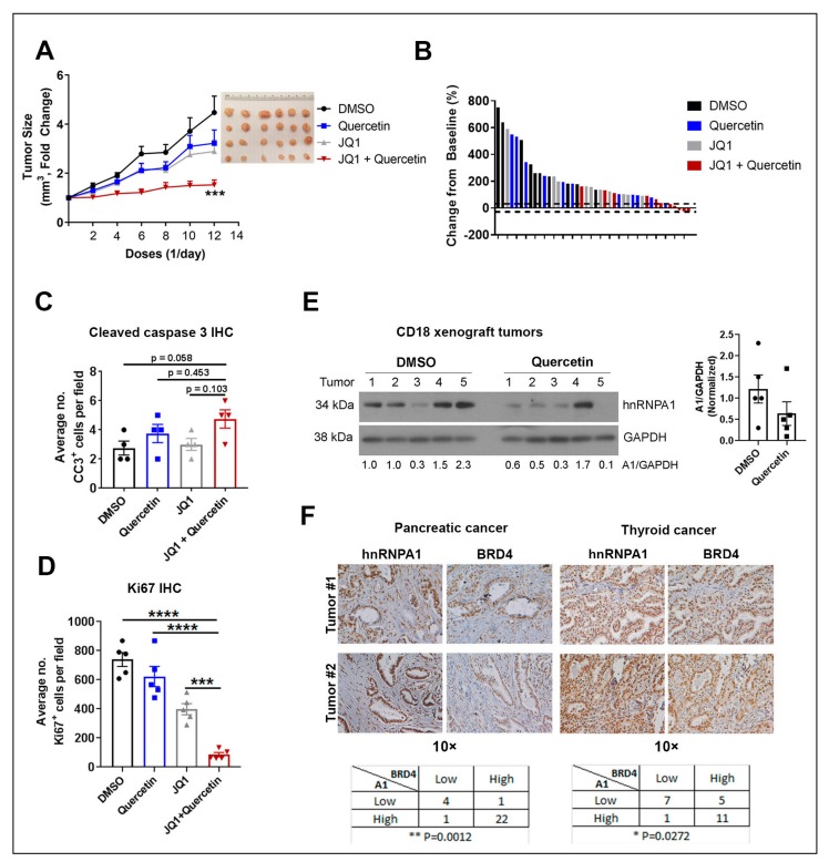
Figure 6.
Quercetin increases the anti-tumor effects of JQ1 in vivo. CD18 pancreatic cancer cells (1.5 × 10⁶) were subcutaneously injected into the flanks of nude mice and allowed to form tumors for 2 weeks. Mice with established tumors were randomized and treated intraperitoneally with DMSO, JQ1 (50 mg/kg), Quercetin (40 mg/kg), or a combination of JQ1 (50 mg/kg) and Quercetin (40 mg/kg) daily Mon–Fri for 3 weeks. (A) Tumor growth was assessed daily Mon–Fri, and tumor volume calculated and normalized to tumor volume at the start of treatment. ***, p < 0.001 compared to the DMSO-treated group. Error bars represent SEM from at least 10 individual tumors. (B) Waterfall plot was generated to demonstrate change in tumor volume relative to start of treatment. Dotted lines indicate 30% cut-off values from the baseline. (C) The tumor specimens were stained for the apoptosis marker cleaved caspase-3 (CC3) by immunohistochemistry and the number of CC3+ cells per 10× field were counted. At least five different sections per individual tumor were counted. Four tumors from each treatment group were used in the analysis. Error bars represent SEM from all tumors analyzed. (D) The tumor specimens were stained for proliferation marker Ki67 by immunohistochemistry and the number of Ki67+ cells per 10× field were counted by ImageJ analysis. At least four different sections per individual tumor were counted. Five tumors from each treatment group were used in the analysis. Two-way ANOVA analysis was performed. ***, p < 0.001 ****, p < 0.0001. Error bars represent SEM from all tumors analyzed. (E) The expression of hnRNPA1 (A1) in CD18 tumors was analyzed by western blot analysis, with GAPDH as loading control. Densitometry graph is shown. Error bars represent SEM from five individual tumors. (F) TMA generated from pancreatic ductal adenocarcinoma (PDAC) (n = 28) and human papillary thyroid cancer (n = 24) specimens were stained for BRD4 and hnRNPA1 by immunohistochemistry, and the relative staining was graded as low (0 or 1+) or high (2+ or 3+). Fisher’s exact test was used to evaluate the relationship between BRD4 and hnRNPA1 (A1) in these specimens. DMSO: dimethyl sulfoxide; JQ1: BET inhibitor; hnRNPA1: heterogeneous nuclear ribonucleoprotein A1; GAPDH: Glyceraldehyde 3-phosphate dehydrogenase (loading control); BRD4: bromodomain-containing protein 4; IHC: immunohistochemistry; TMA: tissue microarray; CD18: human pancreatic cancer cell line.

