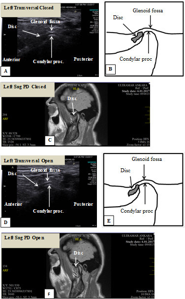
Figure 5.

A 26-year-old female patient referred with complaints of limited mouth opening and pain in left TMJ. Condylar flattening and degenerative changes are observed. (A) Closed mouth ultrasound (transducer placed transversally) image with disc in anterior position; (B) schematic drawing of closed mouth ultrasound (transducer placed transversally) image with disc in anterior position; (C) closed mouth MRI (sagittal plane) image of the patient showing left TMJ with anteriorly positioned disc; (D) open mouth ultrasound (transducer placed transversally) image with disc in anterior position; (E) schematic drawing of open mouth ultrasound (transducer placed transversally) image with disc in anterior position; and (F) open mouth MRI (sagittal plane) image of the patient showing left TMJ with anterior disc displacement without reduction. TMJ, temporomandibular joint.
