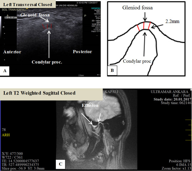
Figure 6.

MRI and ultrasound images of left TMJ of a 55-year-old female patient with joint effusion. The patient was referred with the complaints of severe pain and limitation in mouth opening. (A) Measurement of the distance between mandibular condyle and glenoid fossa on transversal ultrasound image in left closed mouth position; (B) schematic drawing of measurement of the distance between mandibular condyle and glenoid fossa on transversal ultrasound image in left closed mouth position. Measurements higher than 1.76 mm were considered as an increase in synovial fluid thickness leading to joint effusion; (C) sagittal T2 weighted MRI image of the same patient showing joint effusion with high signal intensity anterosuperiorly.
