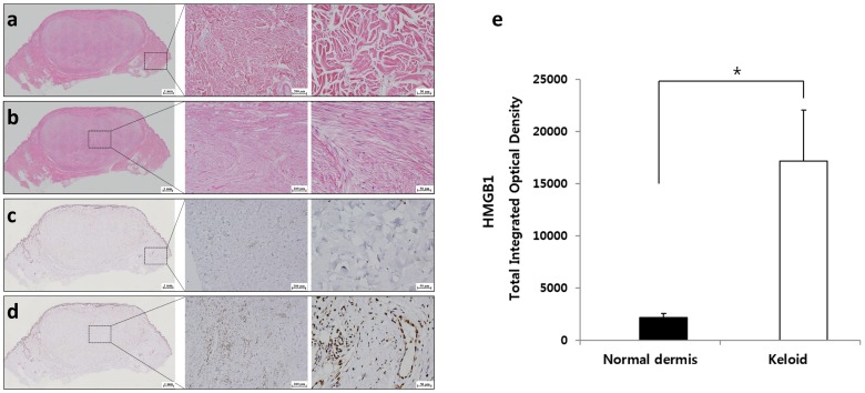
Figure 1.
Histological assessment of keloid tissue. (a) In hematoxylin and eosin (H&E) staining, densely accumulated thick collagen bundles were noted in the keloid tissue. (b) In the normal adjacent dermal tissue, a multidirectional meshwork structure was detected. (c,d) In immunohistochemistry (IHC) of HMGB1, excessively high expression of HMGB1 was noted in the center of the keloid tissue, while expression of HMGB1 was rarely seen in the adjacent normal dermis. (e) Semi-quantitative analysis indicated that the expression of HMGB1 was significantly increased in the keloid tissue compared with that in normal dermal tissue (* p < 0.05, original magnification 100×, 400×).

