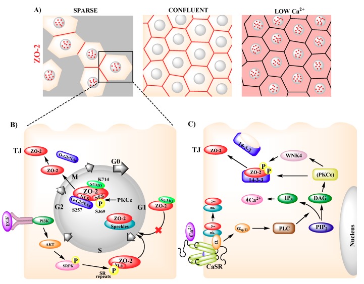
Figure 2.
Intracellular movement of ZO-2. (A) In sparse cultures, ZO-2 is present in nuclear speckles (red dots) and TJs (red cell borders), whereas in confluent cells ZO-2 concentrates at TJs (red cell borders). In cells cultured with low Ca2+ (1–5 μM) ZO-2 distributes diffusely in the cytoplasm (pink cytoplasmic staining) and concentrates in nuclear speckles (red dots). (B) In sparse cultures, epidermal growth factor (EGF) induces AKT activation, which triggers ZO-2 phosphorylation of serine-arginine (SR) repeats by SR protein kinase (SRPK). Nuclear localization signals (NLSs) of ZO-2 and phosphorylated SR repeats induce the arrival of ZO-2 to nuclear speckles at late G1. ZO-2 exit from the nucleus requires the phosphorylation of Ser369 within a nuclear exportation signal (NES) by PKCԑ and the inactivation of bpNLS-2 by O-GlcNAc of Ser257, and is facilitated by SUMOylation of Lys714. (C) Extracellular Ca2+ activates the Ca2+-sensing receptor (CaSR) present in the plasma membrane that signals through the Gαq/11 subunit, activating PLC, which hydrolyzes PIP2 into DAG and IP3. DAG activates PKCԑ, leading to ZO-2 phosphorylation by WNK4. This phosphorylation induces the disassembly of ZO-2 from 14-3-3 proteins in the cytoplasm and triggers the relocation of ZO-2 to the plasma membrane.

