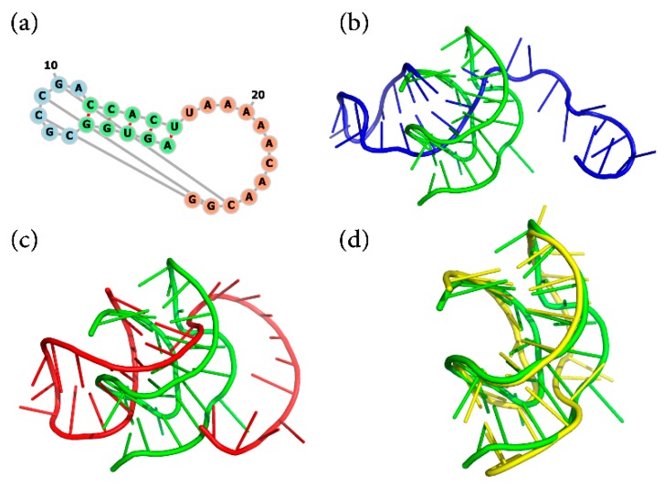
Figure 5.
The secondary structure and predicted 3D structures for an RNA (PDB ID 2AP5): (a) is its secondary structure, (b) is the assembled structure (blue), (c,d) are the optimized structures not using (red) and using (yellow) the pseudoknot interactions as restraints, respectively. The native structure is represented in green cartoon.

