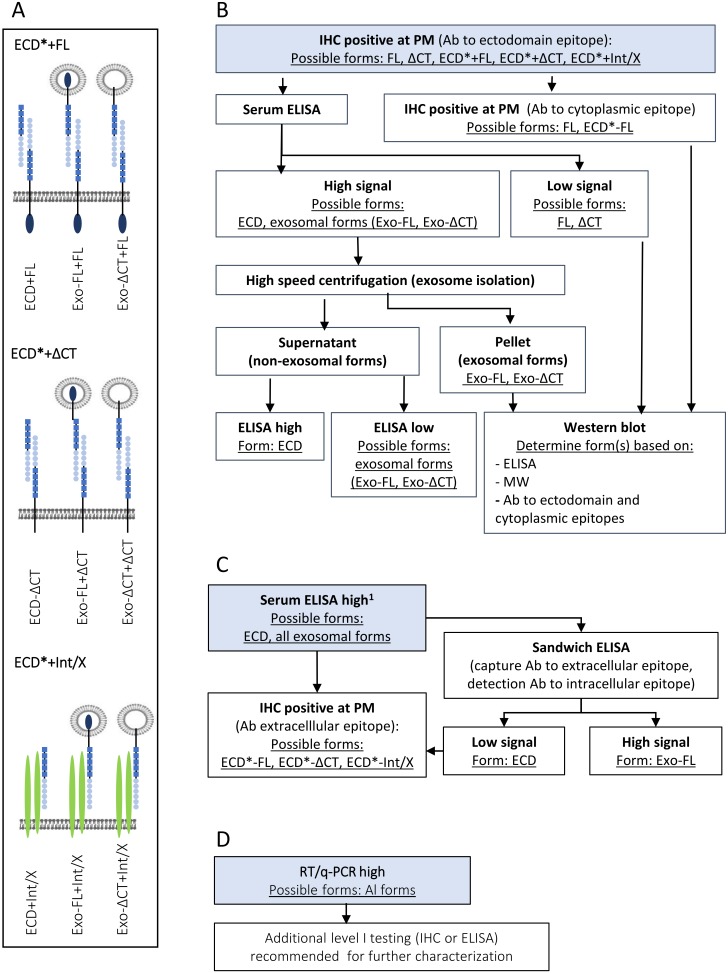
Figure 5.
Suggested algorithm for the assessment of the form and location of L1CAM based on common clinical analyses of L1CAM expression. (A) Analysis of L1CAM is most commonly assessed by IHC using ECD-domain-reacting antibodies. In addition to recognizing L1CAM-FL or L1CAM-ΔCT, positive staining at the plasma membrane can represent homotypic interactions of L1CAM-FL or L1CAM-ΔCT with soluble ECD, or with exosomal forms expressing the ECD (collectively indicated ECD * + FL or ECD * + ΔCT). Positive staining at the plasma membrane may also be due to heterotypic interactions of ECD-containing soluble or exosomal L1CAM forms with other proteins, such as integrins (Int) or other undefined proteins (X). These heterotypic interactions are indicated with ECD * + Int/X. (B) Suggested diagnostic algorithm based on initial positive IHC staining with an ECD-domain-reacting antibody. Suggested follow-up analysis should includes ELISA analysis of serum to determine the level of soluble forms. The presence of FL can be assessed by IHC using a CT-domain-specific antibody, the staining of which should overlap with ECD-specific antibody staining in case of expression of FL. More detailed analyses to distinguish soluble forms can be done by high speed centrifugation of serum samples, by which soluble ECD and exosomal forms can be distinguished. Additional western blot and ELISA analysis using ECD- and CT-reactive antibodies can further stratify exosomal forms by analysis of molecular weight and the presence or absence of either domain. Ab is antibody. Nomenclature of L1CAM forms is also given in the legends of Figure 1C and Figure 2B. (C) Algorithm based on initial assessment of L1CAM in serum by ELISA. ELISA outcome depends on type of ELISA and on the antibodies used. This algorithm assumes direct ELISA, or a sandwich ELISA with either or both antibodies reacting with the ECD, or a commercial assay with proprietary information regarding antibodies. Downstream analyses to distinguish CT-containing forms from FL may include sandwich ELISA combining CT- and ECD-reacting antibodies, and/or IHC with a ECD-reacting antibody, which may be expanded to additional analyses as indicated in B. (D) Increased L1CAM expression as determined by RT/qPCR will not provide information on processing and cleavage form of L1CAM and additional analyses by IHC and ELISA is recommended.

