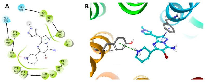
Figure 6.
Docking study of MK-8776 with human P-gp. (A) The generated 2D figure of the docked position of MK-8776 within the P-gp crystal. The polar residues are indicated with cyan bubbles, and the hydrophobic residues are indicated with green bubbles. Furthermore, the cation–π bond in the figure is shown as red arrow, and the π–π interaction is shown as green line. (B) The enlarged 3D figure of the docked position of MK-8776 within the drug-binding site of human P-gp. The structure of MK-8776 is shown as a ball and stick mode with the atoms colored as follows: carbon with cyan, nitrogen with blue, bromine with dark red. The important residues of P-gp are shown as sticks, with the atoms colored as follows: carbon with grey, nitrogen with blue, oxygen with red, hydrogen with white. The cation–π bond is indicated with green dotted line. The π–π stacking interactions are indicated with a blue dotted line.

