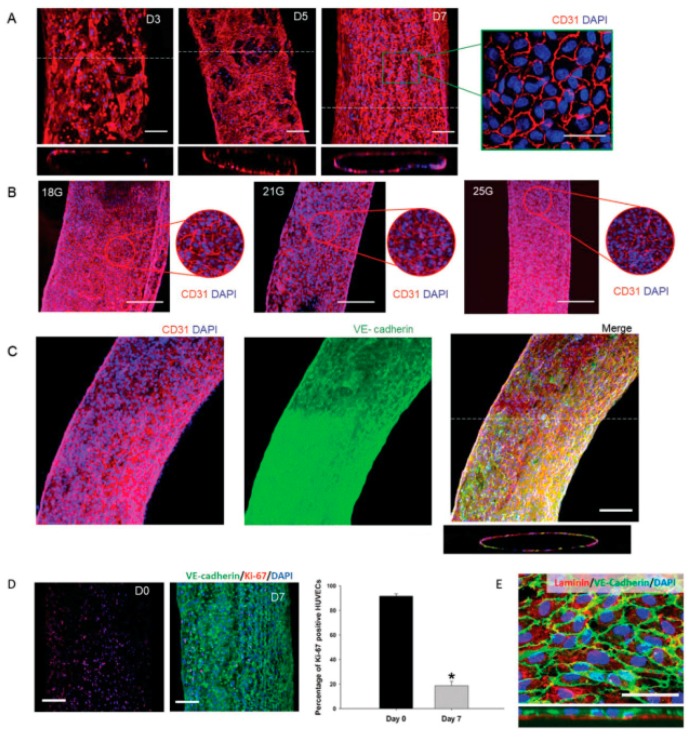
Figure 5.
Formation and maturation of the endothelium after three, five and seven days of culture (scale bars are 100 and 50 µm in the inset) (A). Vessel formation using different needle diameters for bioprinting (scale bar is 200 µm) (B). CD31 and VE-cadherin expression after seven days of culture (scale bar is 100 µm) (C). Ki-67 and VE-cadherin staining demonstrated the decrease of proliferation cells and the consequent vessel stabilization (scale bar is 100 µm. * p < 0.005, N = 3) (D). Detection of laminin in the basolateral side of the endothelial layer (scale bar is 20 µm) (E). Reprinted from Reference [37], with permission from John Wiley and Sons.

