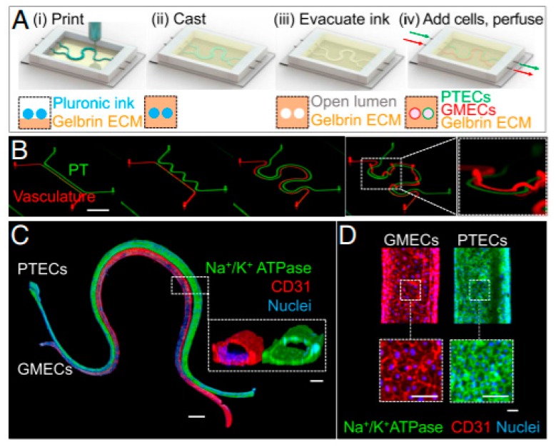
Figure 7.
Schematic illustration of the fabrication of a vascularized 3D bioprinted model (A). Different design patterns ranging from straight to serpentine (Scale bar is 10 mm) (B). Immunostaining of 3D final VasPT model (Scale bar is 1 mm) and cross-section (inset) of the PT and vascular conduits (scale bars are 100 μm) (C). High magnification of the two lumens (scale bars are 100 μm) (D). From Reference [107], with permission from Proceedings of the National Academy of Sciences of the United States of America (PNAS).

