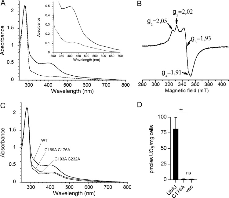
FIG 6.
UbiU-V complex binds two [4Fe-4S] clusters. (A) UV-visible absorption spectra of as-purified UbiU-UbiV (dotted line, 17 μM) and reconstituted holo-UbiU-UbiV (solid line, 15.5 μM). The inset shows an enlargement of the 300- to 700-nm region. (B) X-band EPR spectrum of 339 μM dithionite-reduced holo-UbiU-UbiV. Recording conditions were the following: temperature, 10K; microwave power, 2 mW; modulation amplitude, 0.6 mT. (C) Comparative UV-visible absorption spectra of Cys-to-Ala mutants of UbiU in the UbiU-UbiV complex after metal cluster reconstitution with the following concentrations: 15.5 μM WT, 16.0 μM UbiU C169A C176A, and 16.0 μM UbiU C193A C232A. (A to C) Proteins were in 50 mM Tris-HCl, pH 8.5, 150 mM NaCl, 15% glycerol, 1 mM DTT. (D) UQ8 quantification of ΔubiU cells transformed with pBAD-UbiU (n = 4), pBAD-UbiU C176A (n = 2), or pBAD empty vector (n = 3) and grown overnight in anaerobic SMGN plus 0.02% arabinose. Values are means ± SD. **, P < 0.01; ns, not significant; both by unpaired Student's t test.

