FIG 4.
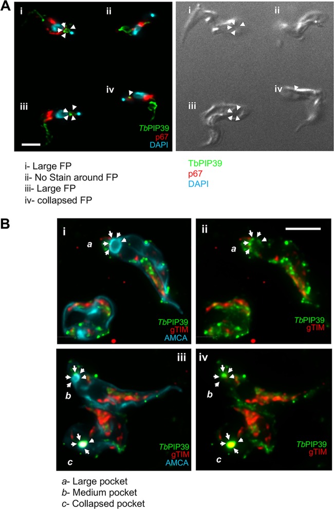
(A) Colabeling of stumpy-form cells (without exposure to cis-aconitate) stained for TbPIP39 (green) and the lysosomal marker p67 (red). The nucleus and kinetoplast are stained in blue. The TbPIP39 labeling is distinct from the lysosome, being positioned unevenly around the flagellar pocket (FP [cell i]) or at a tight focus in cells where the flagellar pocket is collapsed (cells iii and iv). Cell ii has no staining detected at the flagellar pocket region. Bar = 5 μm. (B) Colabeling of stumpy-form cells (without exposure to cis-aconitate) stained for TBPIP39 (green) and the glycosomal gTIM (red). The flagellar pocket and cell surface membrane are labeled blue with aminomethylcoumarin (AMCA). Arrows indicate the distribution of the TbPIP39 signal unevenly around the flagellar pocket (Cells a and b [images i and ii]), or at a tight focus associated with a collapsed flagellar pocket (cell c [images iii and iv]). Bar = 5 μm.

