FIG 6.
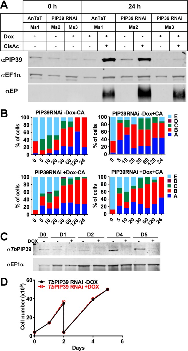
(A) RNAi depletion of TbPIP39. T. brucei EATRO 1125 AnTat1.1 90:13 TbPIP39 RNAi cells or T. brucei EATRO 1125 AnTat1.1 90:13 cells were grown in mice, with or without doxycycline induction. The generated stumpy cells were then exposed, or not, to cis-aconitate to initiate differentiation (with doxycycline remaining in the RNAi-induced samples), and TbPIP39 protein was detected at 24 h. TbPIP39 levels increase during differentiation, but this is greatly reduced in induced RNAi samples. EF1α provides a loading control, this being a little higher in cells exposed to cis-aconitate due to their replication as differentiated procyclic forms. EP procyclin staining shows relative differentiation in the presence or absence of cis-aconitate. (B) Distribution of the glycosomal marker aldolase and the lysosomal marker p67 during differentiation between stumpy and procyclic forms. The panel shows the prevalence of different categories of lysosomal/glycosomal staining (as defined by Herman et al. [23]) at times through differentiation when TbPIP39 was depleted by RNAi or not. The cytological profiles were as follows: A, enlarged lysosomal signal; B, lysosome enlarged but separated into distinct smaller vesicles; C, normal lysosomal size, with glycosomal colocalization; D, no lysosome observed; E, lysosome normal, with no colocalization with glycosomes. (C) Western blot of TbPIP39 in cells differentiated to proliferative procyclic forms with TbPIP39 RNAi induced or not. EF1α shows the loading control. The lower panel shows that by days 4 and 5, the cells remain proliferative (the cells were passaged at day 2) despite expressing significantly less TbPIP39.

