Fig. 1.
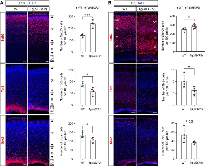
There are more Satb2 positive neurons in Tg(MECP2) FVB mice cortex. Coronal brain sections from E18.5 (a) and P7 (b) Tg(MECP2) FVB mice or WT littermates were stained with cortical neuron marker Satb2, and progenitor markers Sox2 and Tbr2. N ≥ 4. DAPI (blue) was used for nuclear staining. The numbers of positive cells within 100μm bin were counted. Representative sections are shown in left and statistical analysis are shown in right panels, respectively. All statistic data represent means ± SEM. *p < 0.05, ***p < 0.001. Scale bar is 50 μm in E18.5 and 100μm in P7

