Fig. 2.
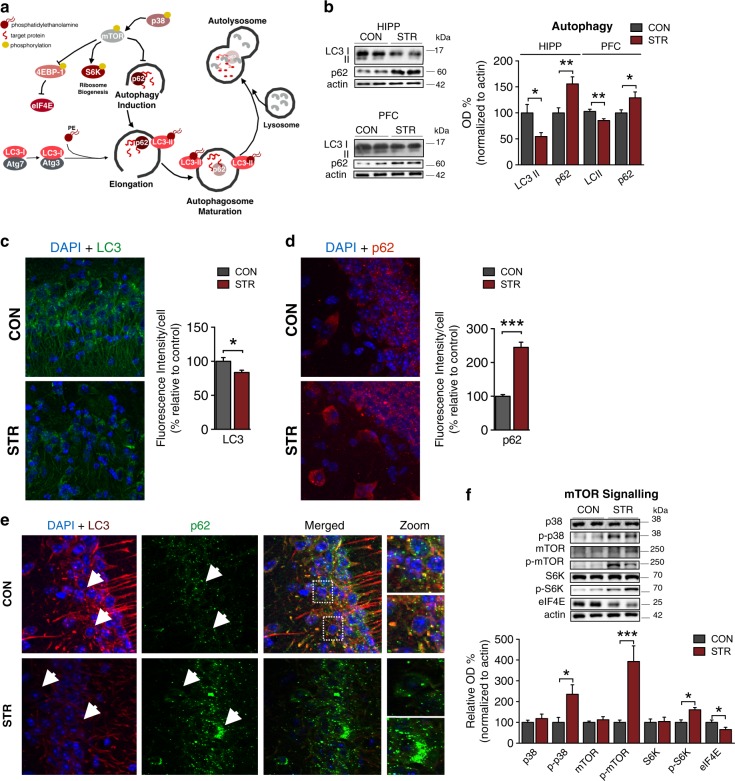
Prolonged exposure to environmental stress inhibits autophagy in a mTOR-dependent manner. a Schematic representation of autophagy highlighting the role of mTOR, LC3, and p62 in this cellular process. b–e Stressed animals exhibited reduced LC3 (PFC: p = 0.003; HIPP: p = 0.026) and increased p62 (PFC: p = 0.029; HIPP: p = 0.004) protein levels as assessed by WB analysis (b); these findings were confirmed by corresponding changes in LC3 (p = 0.044) and p62 (p = 0.044) fluorescence intensity/cell number (c–d) an decrease in their co-localization (e), indicating a stress-driven inhibition of autophagic process. f In line with the above findings, the levels of phosphorylated S6K (p = 0.014), p38 (p = 0.030), and mTOR (p < 0.001) proteins were increased by stress, with decreased levels of eIF4E (p = 0.024), which are indicative of mTOR activation. All numeric data represent mean ± SEM, *p < 0.05; **p < 00.01; ***p < 00.001

