Fig. 3.
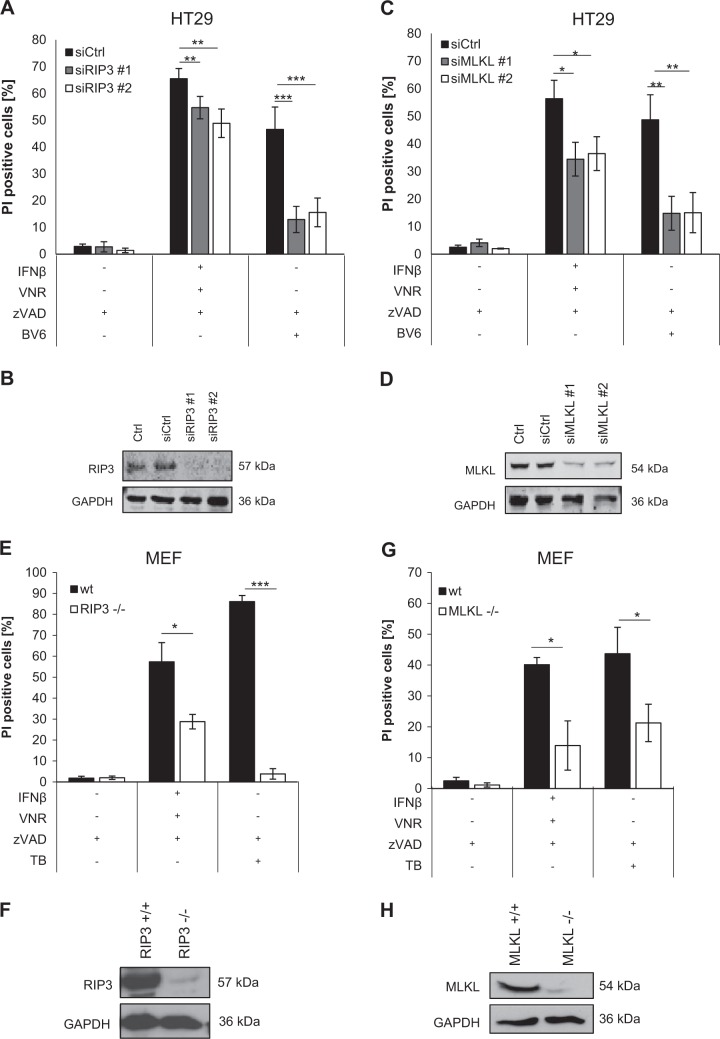
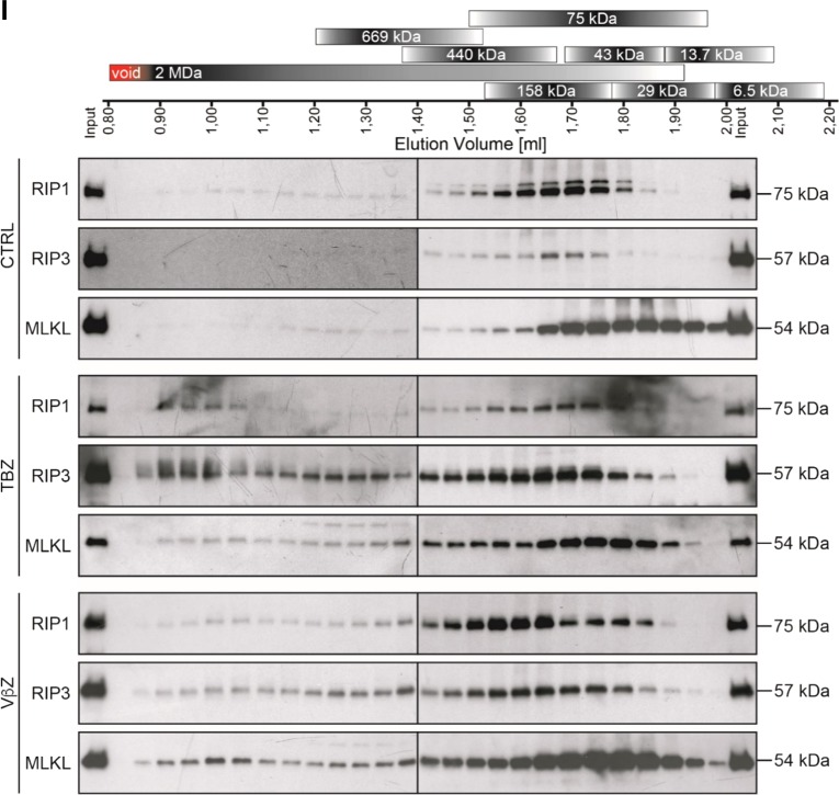
Loss of RIP3 or MLKL inhibits VNR/IFNβ/zVAD.fmk-induced necroptosis. a–d HT29 cells were transiently transfected with siRNA against RIP3 (a, b) or MLKL (c, d) or non-targeting control siRNA (siCtrl). Transfected cells were treated with 10 ng/ml IFNβ, 100 nM VNR, 20 µM zVAD.fmk, and/or 1 µM BV6 for 72 h and cell death was determined by analysis of PI-positive nuclei. Mean and SD of three independent experiments performed in triplicate are shown; *P < 0.05, **P < 0.01, ***P < 0.001 (a, c). Expression of RIP3 and MLKL was assessed by Western blotting, with GAPDH serving as loading control (b, d). e–h Wt MEFs and MEFs deficient for RIP3 or MLKL were treated with 4.5 ng/ml murine IFNβ, 100 nM VNR, 20 µM zVAD.fmk, 10 ng/ml TNFα, and/or 5 µM BV6 for 72 h (IFNβ/VNR/zVAD.fmk) or 5 h (TBZ) and cell death was determined by analysis of PI-positive nuclei. Mean and SD of three independent experiments performed in triplicate are shown; *P < 0.05, ***P < 0.001 (e, g). Expression of RIP3 and MLKL was assessed by Western blotting, with GAPDH serving as loading control (f, h). i MEFs were treated with 4.5 ng/ml murine IFNβ, 100 nM VNR, and 20 µM zVAD.fmk for 72 h or with 10 ng/ml TNFα, 5 µM BV6, and 20 µM zVAD.fmk for 2 h. Lysates were fractionated on a Superose 6 3.2/300 GL column and the resulting fractions, as well as input samples, were analyzed by Western blotting (i). A schematic representation of the calibration, which is shown in detail in Supplementary Figure 4, is depicted above the Western blots


