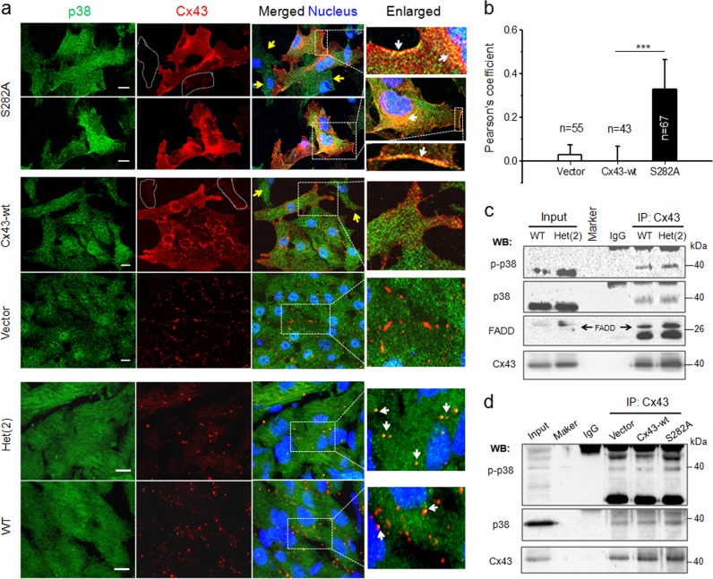
Fig. 8.
Enhanced physical interaction between p38 MAPK and Cx43 in S282A transfected cardiomyocytes and Cx43 S282A+/− hearts. a Representative images of NRVMs transfected with adenovirus and ventricles from WT and Het(2) mice, as indicated, and double-stained with antibodies specific for p38 MAPK (green) and Cx43 (red). The cells indicated with dot irregular shapes and yellow arrows were S282A-negative cells. White arrows point to p38 MAPK colocalized with Cx43 in S282A-positive cell surface and lateral side of the nucleus, and in ventricles of WT and Het(2) mice in enlarged images. Nucleus was stained with Hochest33342, scale bar: 10 μm, n = 3 independent experiments for cell experiment and four hearts for ventricle test in each group. b The quantitative analysis of p38/Cx43 colocalization, represented by Pearson’s correlation coefficient (see Methods), in adenovirus transfected-NRVMs, n = 43–67 cells as indicated, ***P < 0.001, unpaired two-tailed Student’s t test. c Ventricle lysates from WT and Het(2) mice were immunoprecipitated with anti-Cx43 antibody and then Western blotted with anti-p-p38, anti-p38 or anti-FADD antibody, n = 3 independent experiments from 32 mice for each group. d Lysates from vector and Cx43-wt or S282A-transfected cardiomyocytes were immunoprecipitated with anti-Cx43 antibody and then Western blotted with anti-p-p38 or anti-p38 antibody, n = 3 independent experiments for each group

