Fig. 5.
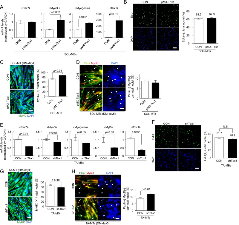
Tbx1 regulates myogenesis in SOL- and TA-muscle-derived myoblasts in vitro. a, e The expression of Pax7, MyoD, Myogenin, Myf5, and Tbx1 in Tbx1-overexpressing (a) or Tbx1-suppressed (e) myoblasts as quantified. Values are presented as mean ± SE (n = 4). b, f Tbx1-overexpressing (b) or Tbx1-suppressed (f) MBs were cultured in growth medium with EdU. The number of EdU(+) cells were counted. Values are presented as mean ± SE (n = 4). Scale bar = 100 µm. c, g Tbx1-overexpressing (c) or Tbx1-suppressed (g) TA-MBs were cultured in differentiation medium for 4 days, and then stained against MyHC (green) and with DAPI (blue). The proportion of MyHC(+) cells among total nuclei was quantified. Values are presented as mean ± SE (n = 3). Scale bar = 100 µm. d, h Tbx1-overexpressing (d) or Tbx1-suppressed (h) TA-MBs were cultured with differentiation medium for 5 days, and then stained for Pax7 (red) and MyoD (green). The number of Pax7(+)MyoD(−) reserve cells were quantified. Values are presented as mean ± SE (n = 3). Scale bar = 100 µm

