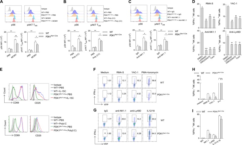
Fig. 5.
PDK1-mTOR signaling plays an important role in NK cell IFN-γ production. a–c The expression levels of phosphorylated S6 and AKT T308 were detected by flow cytometry after splenic lymphocytes were stimulated with MCMV, poly (i:c), or anti-NK1.1 antibody (top) and the absolute MFI were quantified (lower). d Splenic lymphocytes were prepared from poly (i:c)-treated WT mice and pretreated with PI3K-mTOR signaling inhibitors. Intracellular staining was performed to assess the production of IFN-γ. The percentage quantification of IFN-γ-positive NK cells is shown. e The expression levels of the early activation markers CD69 and CD25 were detected by flow cytometry after NK cells were stimulated with IL-15 or Poly(i:c) in the indicated mice. f–i Representative flow cytometric profiles (f, g) and the percentage (h, i) of IFN- γ production in splenic NK cells from indicated mice following stimulation with tumor cells (f, h), plate-coated antibodies or cytokines (g, i). The data represent the mean ± s.d and are representative of three independent experiments

