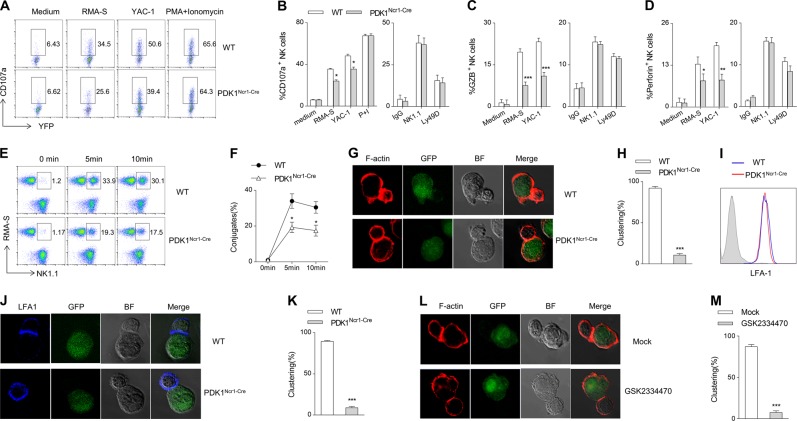
Fig. 6.
PDK1 regulates NK cell conjugation with cellular targets. a–d Representative flow cytometric profiles (a) and the percentage of CD107a (b), granzyme B (c) and perforin (d) positive NK cells from indicated mice following stimulation with tumor cells or plate-coated antibodies. e, f The percentage of NK-target cells conjugate formation from the indicated mice. g, j NK cells from the indicated mice were mixed with RMA-S cells to analyze the localization of F-actin (g) or LFA-1 (j) in the conjugates. h, k The data are expressed as the percentages of conjugates with the polarized accumulation of F-actin (h) or LFA-1 (k). i The expression levels of LFA-1 in WT or PDK1-deficient NK cells was detected by flow cytometry. l The expanded WT NK cells were pretreated with GSK2334470 and then mixed with RMA-S cells for 10 min to analyze the localization of F-actin in the conjugates. m The data are expressed as the percentage of conjugates with the polarized accumulation of F-actin. The data represent the mean ± s.d and are representative of three independent experiments

