Fig. 1.
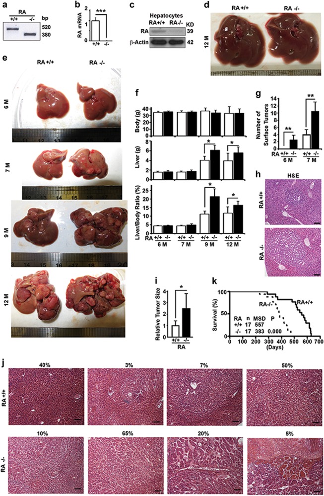
RASSF1A suppresses hepatocarcinogenesis and promotes survival in DEN-treated mice. a PCR analysis of DNA samples from mouse tails to genotype wild-type (RA+/+) and RASSF1A−/− mice (RA−/−). b Quantitative real-time PCR analysis of the levels of RASSF1A mRNA in liver tissues from wild-type and RASSF1A−/− mice. c Representative immunoblot showing levels of RASSF1A protein in hepatocytes isolated from wild-type and RASSF1A−/− mice. d Representative images of liver tissues from 12-month-old untreated wild-type and RASSF1A−/− mice in normal conditions. e Representative images of liver tissues from DEN-treated wild-type and RASSF1A−/− mice at different ages. f Plots of body weights, liver weights, ratios of body weight to liver weight of mice as shown in e. g Plots of number of surface tumors of mice as shown in e. h Comparative H&E staining among the liver tissues from DEN-treated 6-month-old mice described in e. Bar = 20 µm. i Plots of tumor size as shown in h. j Representative images showing different types of H&E staining of liver tissues from DEN-treated 12-month-old mice as shown in e. Bar = 20 µm. The percentage shown on top of each panel is the relative frequency of the type. k The Kaplan–Meier survival curves showing the survival times of male littermates of wild-type and RASSF1A−/− mice treated with DEN. n number of mice, MSD median survival days. The significance of difference between two groups was estimated by log-rank test and p value was the probability larger than the χ2 value. Here and later, all experiments were repeated at least three times. ns, not significant or p > 0.05; *p ≤ 0.05; **p ≤ 0.01; and ***p ≤ 0.001

