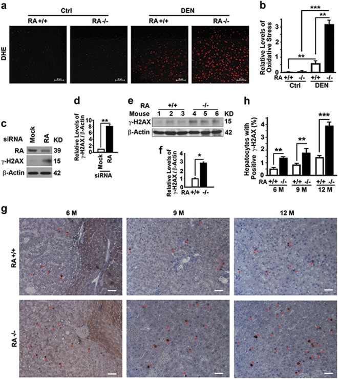
Fig. 2.
RASSF1A suppresses oxidative stress and DNA damage in mouse liver tissues. a, b Representative images (a) and quantification (b) showing levels of oxidative stress revealed by dihydroethidine hydrochloride (DHE) staining in liver tissues from wild-type and RASSF1A−/− mice treated with vehicle (Ctrl) or DEN for two days. Bar = 50 µm. c, d Representative immunoblot (c) and quantification (d) showing γ-H2AX levels in HeLa cells treated with random (Mock) or RASSF1A-specific siRNA (RA). e, f Representative immunoblot (e) and quantification (f) showing γ-H2AX levels in liver tissues from DEN-treated 6-month-old wild-type and RASSF1A−/− mice. g Representative immunostaining of γ-H2AX in liver tissue sections from DEN-treated wild-type and RASSF1A−/− mice as described in (Fig. 1e). Red arrows indicate γ-H2AX positive cells. Bar = 10 µm. h Plots of percentage of γ-H2AX positive cells to total cells in liver sections as shown in g

