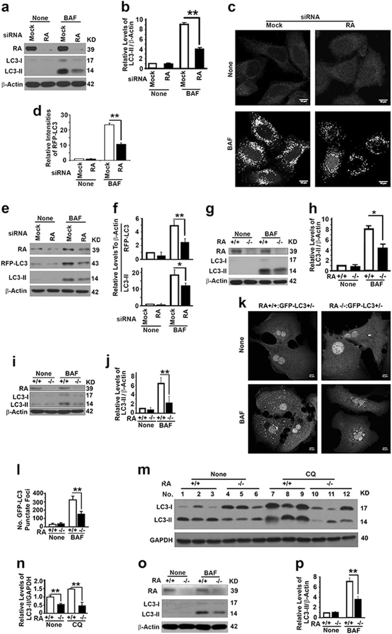
Fig. 3.
RASSF1A activates autophagy flux. a, b Representative immunoblot (a) and quantification (b) showing LC3-II levels in HeLa cells treated with random (Mock) or RASSF1A-specific siRNAs (RA) in the absence (None) or presence of lysosomal inhibitor BAF (10 µM overnight before harvest). c, d Representative images (c) and quantification (d) showing the number of RFP-LC3 punctate foci in HeLa cells stably expressing RFP-LC3 treated with random (Mock) or RASSF1A-specific siRNAs (RA) in the absence (None) or presence of BAF. Bar = 10 µM. e, f Representative immunoblots (e) and quantification showing the levels of exogenous RFP-LC3 and endogenous LC3-II (f) in similar cells as shown in c. g, h Representative immunoblot (g) and quantification (h) showing LC3-II levels in wild-type and RASSF1A−/− MEFs in the absence (None) or presence of BAF. I, j Representative immunoblot (i) and quantification (j) showing LC3-II levels in hepatocytes similarly isolated from mice shown in g. k, l Representative images (k) and quantification (l) showing the number of GFP-LC3 punctate foci in hepatocytes isolated from GFP-LC3 transgenic wild-type and RASSF1A−/− mice in the absence (None) or presence of BAF. Scale bar, 10 µM. m, n Representative immunoblot (m) and quantification (n) showing LC3-II levels in liver tissues from wild-type and RASSF1A−/− mice injected with saline (None) or lysosomal inhibitor CQ. o, p Representative immunoblot (o) and quantification (p) showing LC3-II in hepatocytes isolated from 4-month old DEN-treated wild-type and RASSF1A−/− mice in the absence or presence of BAF

