Fig. 1.
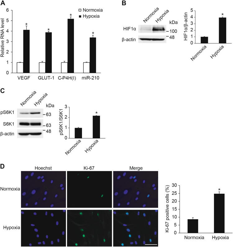
Hypoxia promotes VSMC proliferation through the mTOR signaling pathway. a PASMCs were exposed to normoxia or hypoxia for 24 h and subjected to qRT-PCR analysis of VEGF, GLUT-1, C-P4H(I) and miR-210. The relative levels of mRNA expression normalized to 18S rRNA were quantitated. The expression levels of miR-210 were normalized to U6 snRNA. *p < 0.05. b Total cell lysates from PASMCs exposed to normoxia or hypoxia for 24 h were subjected to immunoblot analysis with anti-HIF1α or anti-β-actin antibody. By densitometry, relative amounts of HIF1α protein normalized to β-actin were quantitated. *p < 0.05. c Total cell lysates from PASMCs exposed to normoxia or hypoxia for 24 h were subjected to immunoblot analysis with antibody against pS6K1, S6K1 or β-actin. By densitometry, relative amounts of phosphorylated S6K1 protein normalized to total S6K1 were quantitated. *p < 0.05 d Representative microphotographs of Ki-67 immunostaining of PASMCs exposed to normoxia or hypoxia, and calculation of Ki-67 index. Approximately 200 cells from at least 10 independent fields were counted for each condition, and Ki-67-positive cells are presented as a percentage of the total population. Scale bar represents 50 μm. *p < 0.05

