Fig. 3.
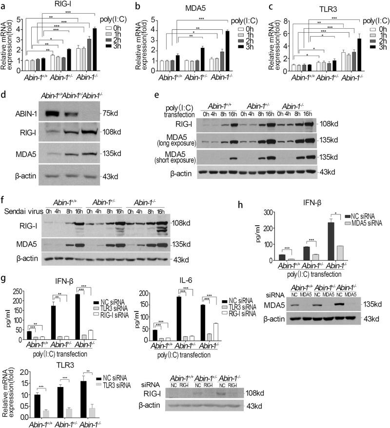
ABIN-1 deficiency increases basal and poly (I:C)/virus-induced TLR3, RIG-I, and MDA5 expression. a–c qPCR assay of RIG-I (a), MDA5 (b), and TLR3 (c) expression upon 2 μg/ml poly(I:C) transfection for 0–3 h in Abin-1+/+, Abin-1+/−, and Abin-1−/− MEFs. d Western blot analysis of the basal levels of ABIN-1, RIG-I, and MDA5 in Abin-1+/+, Abin-1+/−, and Abin-1−/− MEFs. e Western blot analysis of poly(I:C) transfection-induced RIG-I and MDA5 expression at the indicated time points (0, 4, 8, and 16 h). Poly(I:C), 2 μg/ml. f Western blot analysis of Sendai virus-induced RIG-I and MDA5 expression at different time points (0-16 h). g Effect of TLR3 or RIG-I knockdown on poly(I:C)-induced IFN-β and IL-6 production. MEFs were transfected with 50 nM negative control (NC)/RIG-I/TLR3 siRNA for 48 h and then transfected with 2 μg/ml poly(I:C) for 4.5 h, followed by collection of supernatants for ELISA. The lower two graphs are knockdown efficiency of TLR3 and RIG-I. h Effect of MDA5 knockdown on poly(I:C)-induced IFN-β production. Procedures are the same as described in g. The lower graph is the MDA5 knockdown efficiency. Data are presented as the mean ± S.D. from at least three independent experiments. *P < 0.05, **P < 0.01, ***P < 0.001

