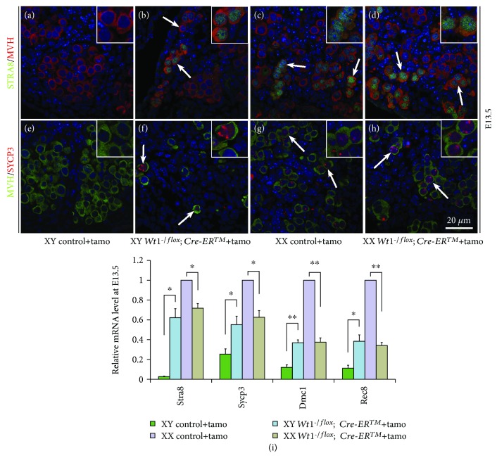
Figure 5.
The meiotic genes were expressed in germ cells of both male and female Wt1-/flox; Cre-ERTM mice after tamoxifen induction. Wt1flox/flox females were crossed with Wt1-/flox; Cre-ERTM mice, and the pregnant females were injected with tamoxifen at E9.5 to induce Cre activity. Wt1flox/flox and Wt1-/flox embryos were used as controls. A–H: immunofluorescence analysis of STRA8/MVH (A–D) and SYCP3/MVH (E–H) in control and Wt1-/flox; Cre-ERTM embryos at E13.5. Germ cells were labeled with MVH. DAPI (blue) was used to stain the nuclei. The arrows point to double-positive germ cells. I: real-time PCR analysis of Stra8, Sycp3, Dmc1, and Rec8 in control and Wt1-/flox; Cre-ERTM embryos at E13.5. Hprt1 was used as an endogenous control. The data are presented as mean ± SEM. ∗P < 0.05; ∗∗P < 0.01.

