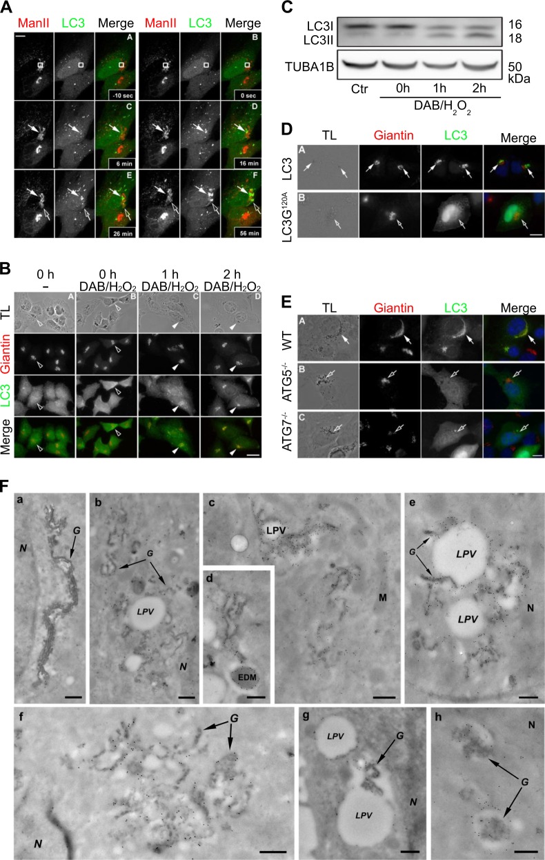
Fig. 1.
LC3 recruitment to wounded or intoxicated Golgi apparatus (GA). a Human cervix carcinoma HeLa cells stably transduced with GFP-LC3 (LC3) were transfected with pManII-mCherry (ManII). Photodamage was applied on a confocal microscope equipped with a 2-Photon Laser. Pictures show cells 10 seconds before photodamage A, after photodamage with the rectangle indicating the photodamaged zone B and representative images of cells at several time points after photodamage C–F. White arrows follow the photodamaged zone showing early LC3 recruitment. Empty arrows indicate appearance of late LC3 recruitment elsewhere in the GA. B HeLa cells stably co-expressing ManII-HRP and GFP-LC3 were fixed directly A or were treated with DAB-H2O2 mix for 30 min at 4 °C then either fixed B or incubated in fresh complete medium for 1 or 2 h before fixation C, D. Cells were subsequently immunostained with anti-Giantin antibodies (Giantin). White arrowheads indicated recruitment of LC3 to the damaged GA. Empty arrowheads indicate absence of LC3 in the Golgi apparatus surroundings. Transmitted light (TL) images were taken to detect the DAB precipitates. Scale bars equal 10 μm. c HeLa ManII-HRP cells without prior treatment or upon treatment with DAB-H2O2 mix for 30 min at 4 °C then were either lysed directly or incubated in fresh complete medium for different time points prior to lysis. Lysates were separated by SDS-PAGE, and then electrotransferred onto a nitrocellulose membrane for immunodetection with anti-LC3 and anti-tubulin antibodies. d HeLa cells expressing ManII-HRP were transiently transfected with GFP-LC3 wt a or GFP-LC3 G120A mutant plasmids b. Cells were treated with DAB-H2O2, fixed 2 h after wash-out, and immunostained with anti-Giantin antibodies. e Wildtype, ATG5−/− or ATG7−/− mouse embryonic fibroblasts (MEFs) were co-transfected with pManII-HRP and pEGFP-LC3 wt plasmids 24 h before the experiment. Cells were treated with DAB-H2O2, fixed 2 h after wash-out and stained with anti-Giantin antibodies. White arrows indicate colocalization of LC3 with Giantin and DAB precipitate. Empty arrows indicate absence of LC3 at the GA surrounding. Transmitted light (TL) images were taken to detect the DAB precipitates. Scale bars equal 10 μm. f HeLa ManII-HRP GFP-LC3 were treated with DAB-H2O2 for 30 min at 4 °C then incubated in fresh complete medium for 0, 2, or 4 h before processing for immuno-EM. GFP-LC3 was detected using antibodies against the GFP tag and secondary antibodies labeled with 10 nm gold particles. Representative images are depicted for 0 a, 2 b–d, and 4 h after DAB-H2O2 treatment e–h. LPV = LC3-positive vacuoles; G = Golgi; N = nucleus; M = mitochondria. Scale bars equal 300 nm

