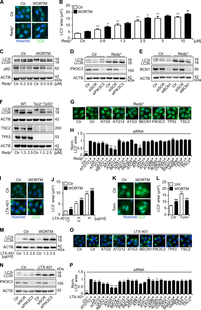
Fig. 7.
LC3 aggregation is independent of PIK3C3 and Beclin-1 but dependent on ATG5, ATG12 and ATG3. a–h Human osteosarcoma U2OS wildtype or stably GFP-LC3 expressing cells were pre-treated with wortmannin (WORTM; 1 µM) or submitted to the RNA interference-mediated silencing of PIK3C3 and BCN1 followed by photodynamic therapy (PDT) with redaporfin (Redp*) a–e LTX-401 i, j, and m, n and torin k, l. Six hours later, LC3 aggregation was evaluated by microscopy in GFP-LC3 expressing cells whereas WT cells were processed for immunoblotting for the assessment of LC3 lipidation. MEFs WT or MEFs DKO for TSC2 and TP53 were also submitted to redp-PDT followed by the assessment of LC3 lipidation by means of immunoblotting f. Alongside, U2OS cells expressing GFP-LC3 cells were submitted to RNA interference-mediated silencing of 24 different autophagy-related genes followed by redp* g, h or LTX-401 o, p treatment. Six hours later, GFP-LC3+ puncta were evaluated by fluorescence microscopy. Representative images of GFP-LC3+ puncta are depicted for redp* a, g; LTX-401 i, o; or torin k and the quantitative analysis that shows the average area of GFP-LC3+ puncta is presented in b, h; j, p; and l, respectively. Representative western blots of LC3 lipidation are depicted in c–f for redp* and in m, n for LTX-401. Data are presented as means ± SEM of at least two independent experiments. (Two-way ANOVA, ***p < 0.001 versus untreated cells; #p < 0.5, ##p < 0.01, ###p < 0.001 versus the presence of wortm or PIK3C3 or BCN1 silencing. For h, p, two-way ANOVA, ***p < 0.001 versus treated cells in the presence of the siCTR). Size bar equals 10 µm

