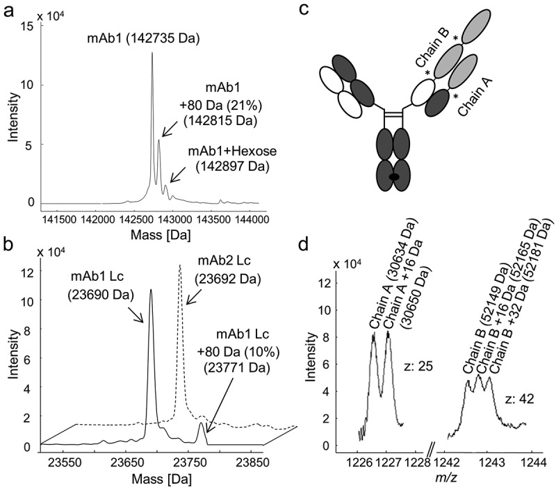
Figure 1.

Intact and reduced mass analysis of human IgG1s mAb1 and mAb2, and bispecific IgG-fusion protein BsAbA expressed in Chinese hamster ovary cells. Deconvoluted mass spectra of (a) the N-deglycosylated mAb1, and (b) the reduced light chain (Lc) of mAb1 (solid line) and mAb1-related antibody mAb2 (dotted line). (c) Schematic illustration of BsAbA consisting of four different chains and based on the CrossMabCH1-CL format including three 4–1BB ligand domains (light grey) allocated to chain A and B. * denotes glycine-serine linkers. (d) Mass spectrum of the N-deglycosylated and reduced BsAbA (chain A, charge state z: 25; chain B, charge state z: 42).
