Figure 1.
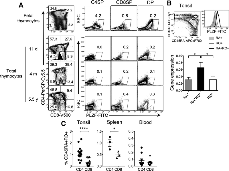
PLZF+CD4 T cells express both CD45RA and CD45RO and reside in lymphoid tissues. (A) Total cells from fetal or child thymii samples were stained with surface antibodies and gated based on CD4 and CD8 expression. PLZF+ cells in each subset are shown. PLZF-expressing NKT cells were excluded from our analyses by CD1d-Tetramer staining (Supporting Information Fig. 2). One representative is shown of total 3 fetal thymi, which were examined in independent experiments. Total 9 after-birth thymi were examined in independent experiments of which, three thymi of different ages are shown. d, m, and y indicates days, months, and years, respectively of subject’s age. (B) Left panel, Tonsillar lymphocytes were stained with surface antibodies, followed by intranuclear staining for PLZF. CD4+ T cells were gated into CD45RA+, CD45RO+, and CD45RA+RO+ cells were compared for PLZF expression by flow cytometry (n = 8) from six independent experiments. Right panel, CD45RA+, CD45RO+ and CD45RA+RO+CD4 T cells from tonsils were sorted to prepare RNA and then cDNA. PLZF gene expression was compared by qPCR. Relative expression of PLZF over HPRT is shown. Error bars represent the mean ± SEM from triplicate wells. *p < 0.05. Two independent experiments were performed. (C) CD4 and CD8 T cells from tonsils (n = 17), spleens (n = 3), and blood (n = 10) were analyzed by flowcytometry as in (B) to compare the frequency of CD45RA+RO+CD4 versus CD8 T cells. Error bars represent the mean ± SEM from pooled data from at least three independent experiments. *p < 0.05; ****p < 0.0001 (t-test).

