Figure 2.
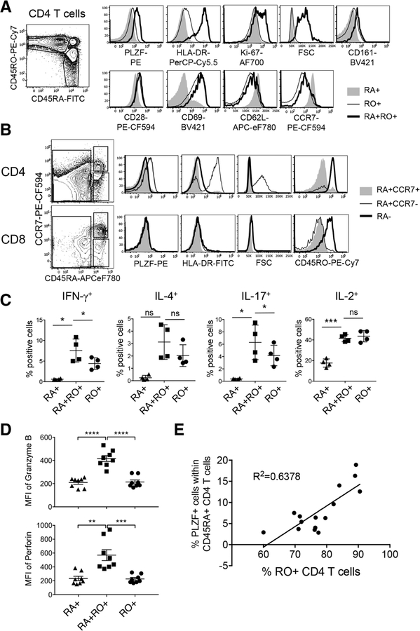
PLZF+CD45RA+RO+CD4 T cells show characteristics of terminally differentiated effector memory cells (A) CD45RA+, CD45RO+, CD45RA+RO+CD4 T cells from tonsils were analyzed for the expression of indicated molecules by flow cytometry after surface and intranuclear staining. Total 16 tonsils were examined and one representative data are shown. (B) Naїve (RA+CCR7+), central and effector memory (RA−) and Temra (RA+CCR7−) CD4 (top) or CD8 (bottom) T cells were assessed for the expression of indicated molecules by flow cytometry. Total 16 tonsils were examined and one representative data are shown. (C) Tonsillar CD4 T cells were stimulated with PMA and Ionomycin for 5 hours in the presence of Monensin, followed by intracellular staining to assess the expression of cytokines. Graphs illustrate the summary of data as mean ± SEM (n = 4). (D) Freshly isolated lymphocytes from tonsils were gated as CD45RA+, CD45RO+, and CD45RA+RO+CD4 T cells and assessed for the expression of Granzyme B and perforin after staining with cocktail of antibodies. Error bars represent the mean ± SEM. ns, not significant. (n = 8) (E)± Tonsillar CD4 T cells were examined by flow cytometry to assess the frequency of PLZF+ cells. The frequency of PLZF+ cells within CD45RA+CD4 T cells was calculated and plotted against the frequency of CD45RO+CD4 T cells (n = 15). ns, not significant; *p < 0.05; **p < 0.01; ***p < 0.001; ****p < 0.0001 (t-test). All data are representative of or pooled from at least three independent experiments.

