. 2019 Sep 17;8:e47155. doi: 10.7554/eLife.47155

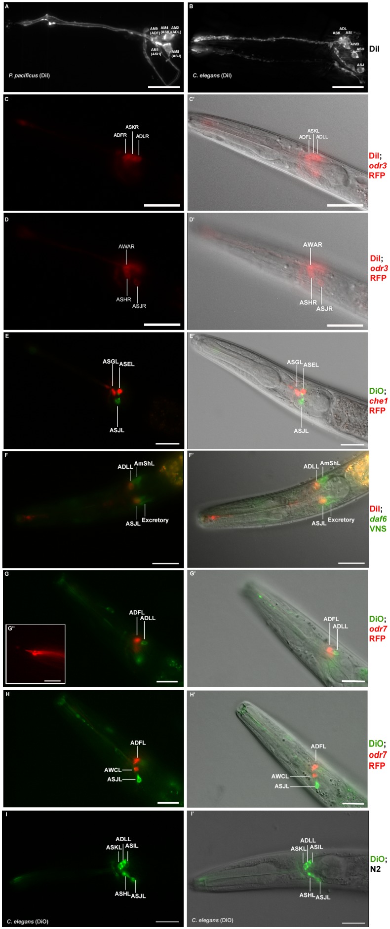
Figure 4. Dye filling and reporter gene expression in P. pacificus amphid neurons.

(A) Stacked and deconvoluted fluorescent images of DiI stained amphid neurons in a P. pacificus young adult hermaphrodite (left lateral view). AM9(ADF), AM4(ASK), AM2(ADL), AM1(ASH), and AM8(ASJ) amphid neurons stain robustly. (B) In C. elegans, DiI stains the ASK, ADL, ASI, AWB, ASH, and ASJ neurons. (C–H) Single plane fluorescent images in P. pacificus. (C–D) DiI stained Ppa-odr-3p::rfp transgenic J3 larvae showing the three dorsal amphid neurons and the larger ASH and ASJ neurons in two focal planes. (E) Ppa-che-1p::rfp transgenic J4 hermaphrodite showing expression in ASE and ASG. (F) A DiI stained Ppa-daf-6p::venus J4 larva showing ADL and ASJ staining, anterior to the amphid sheath and the excretory cells, respectively. (G, H) A DiO stained Ppa-odr-7p::rfp young adult in two focal planes showing dye filling in ADL and ASJ, and RFP expression in ADF and AWC. (G’ inset) The dendritic ends of another Ppa-odr-7p::rfp adult show the prominent PCMC of ADF with double cilia (bottom), and the smaller PCMC of ASK with a single cilium. (I) A DiO stained C. elegans young adult showing five dye filled neurons ASK, ADL, ASI, ASH, and ASJ. Scale bar: 5 µm. (C’–I’) DIC overlay of the same DiI fluorescence images. Anterior is left and dorsal is up. Scale bar: 20 µm. (Representative images based on: Ppa-odr-3p::rfp n = 22; Ppa-che-1p::rfp n = 31; Ppa-daf-6p::venus n = 60; PS312 n = 10; Ppa-odr-7p::venus n = 26). Additional Z-stacks are available: Figure 4—figure supplements 15 and Figure 1—videos 13.

Figure 4.

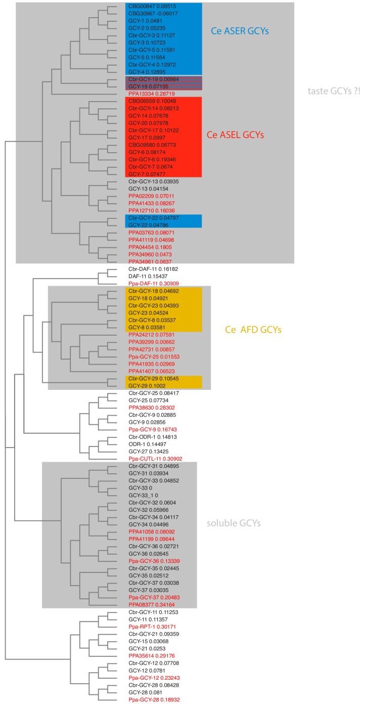
Figure 4—figure supplement 1. Receptor-type guanylyl cyclases in P. pacificus (red font) and Caenorhabditis species (black font).

Figure 4—figure supplement 1.

Figure 4—figure supplement 2. Abbreviated phylogeny of ASE taste receptor-type guanylyl cyclases in P. pacificus (red font) and Caenorhabditis species (black font).
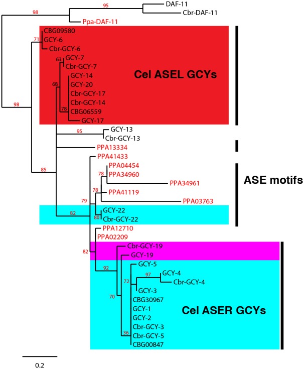
Figure 4—figure supplement 2.

DAF-11 orthologs form an outgroup. Only branch support ≥30% is shown.
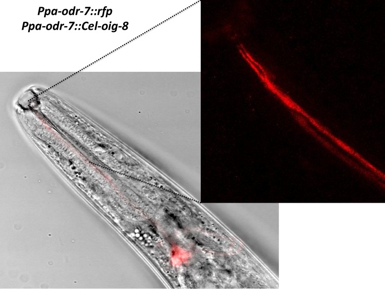
Figure 4—figure supplement 3. Ppa-odr-7p::Cel-oig-8 mis-expression.

Figure 4—figure supplement 3.

An overlay of the AM7(AWC) cell body showing no change in the ciliated endings of AM7 and AM9(ADL)(blowup panel).
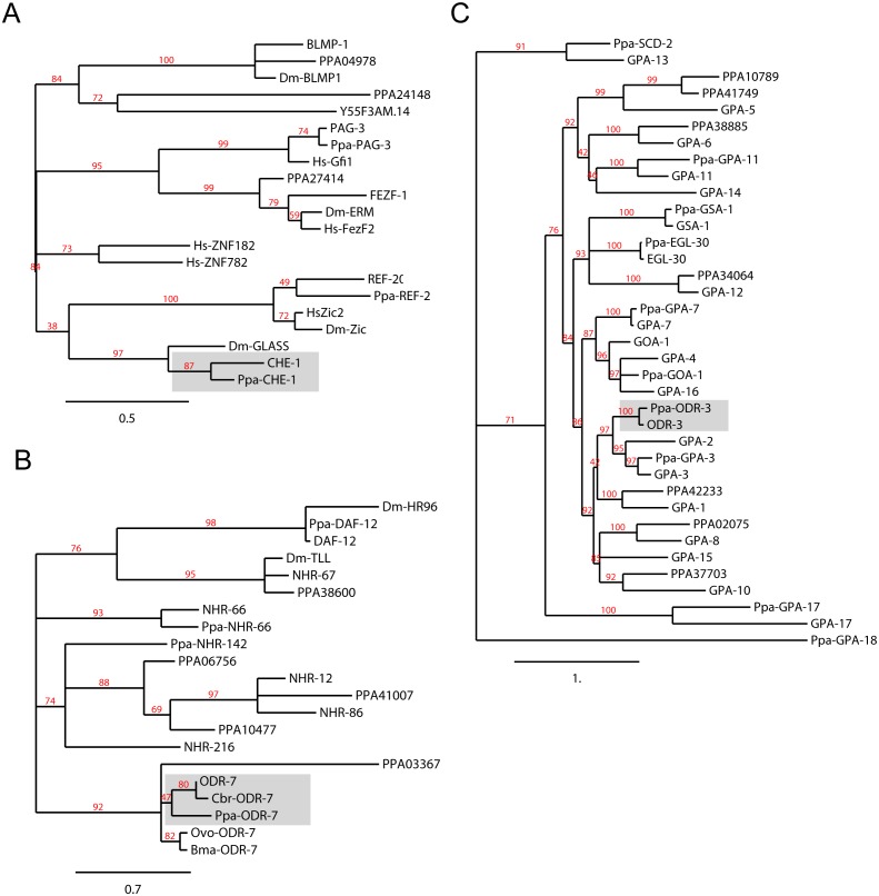
Figure 4—figure supplement 4. Orthology assignments for che-1, odr-7 and odr-3.

Figure 4—figure supplement 4.

Phylogenetic trees for (A) the CHE-1 Zinc finger transcription factor, (B) the ODR-7 nuclear hormone receptor, and (C) the ODR-3 G-alpha protein subunits. The C. elegans and P. pacificus orthologs are shaded. Maximum Likelihood phylogeny trees were generated by PhyML (phylogeny.fr). For the nuclear hormone receptor tree building, only the DNA binding C4 domain was used. Midpoint rooting was used and only branch support ≥30% is shown. Protein sequences for P. pacificus are ‘Ppa-’ (orthology same as Wormbase.org), or ‘PPA’ (no orthology assignment), whereas the rest are C. elegans names. Sequences from additional species are Drosophila melanogaster (Dm), Homo sapiens (Hs), Caenorhabditis briggsae (Cbr), Brugia malayi (Bma), and Onchocerca volvulus (Ovo).
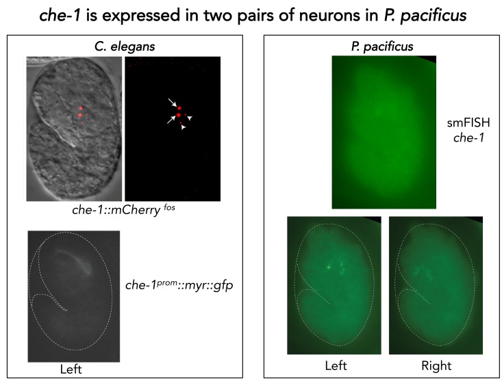
Figure 4—figure supplement 5. Single molecule FISH of Ppa-che-1.

Figure 4—figure supplement 5.

(Left) Representative images of C. elegans embryos carrying two different Cel-che-1 reporters: a translational fusion of CHE-1 to mCherry in the context of a recombineered fosmid construct shows the expected nuclear localization in a pair of neurons identified as the ASE neurons (arrows) as well as two apoptotic bodies (arrow heads) corresponding to the dying ASE-sister cells. A 1.3 Kb fragment upstream of the Cel-che-1 coding sequence also drives expression in the ASE neurons, in this case visualized with a myristoylated GFP reporter. (Right) A representative P. pacificus embryo stained with the smFISH probe set against Ppa-che-1. Individual planes focused on the left or right sides of the embryo show two neurons on each side.
Figure 4—video 1. Z-stack images of DiI stained J3 larva (ventral view).
Download video file (122.4KB, mp4)
DOI: 10.7554/eLife.47155.037
Anterior is up.
Figure 4—video 2. Z-stack images of Ppa-che-1p::rfp stained with DiO.
Download video file (52.1KB, mp4)
DOI: 10.7554/eLife.47155.038
Anterior is to the left and dorsal is to the right.
Figure 4—video 3. Z-stack images of Ppa-odr-7p::rfp.
Download video file (1.6MB, mp4)
DOI: 10.7554/eLife.47155.039
Anterior is to the left and dorsal is to the right.