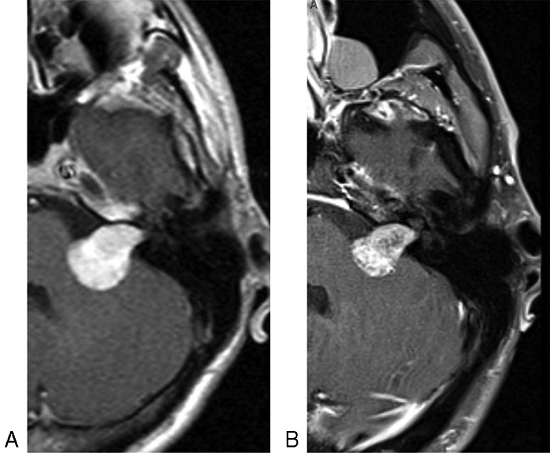
Fig. 2.

Axial T1-weighted postgadolinium magnetic resonance imaging demonstrating left vestibular schwannoma extending into the cerebellopontine angle prior to bevacizumab therapy ( A ) and at the end of bevacizumab therapy ( B ). Tumor volume at the end of bevacizumab was 49.5% of the starting volume.
