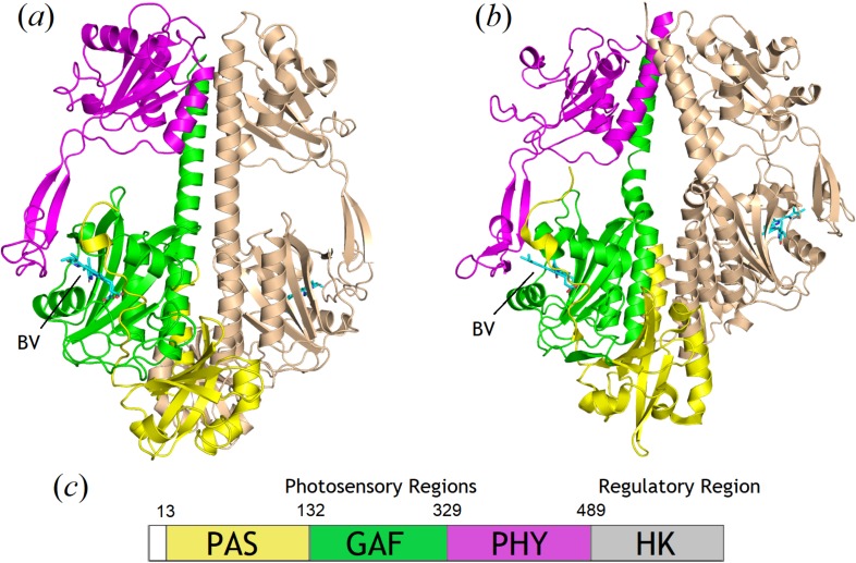
FIG. 1.
Comparisons of the SaBphP2 PCM (a) in the wild-type and SaBphP1 PCM (b) in the wild-type forms. The PAS, GAF, and PHY domains are colored yellow, green, and magenta, respectively. The PCM is a dimer with one monomer highlighted in gold. The kink at the helical transition from GAF to PHY is apparent in panel (b) and BV is marked in panels (a) and (b). (c) Schematic presentation of domain organization of BphPs is below with sequence numbers provided for SaBphP2.

