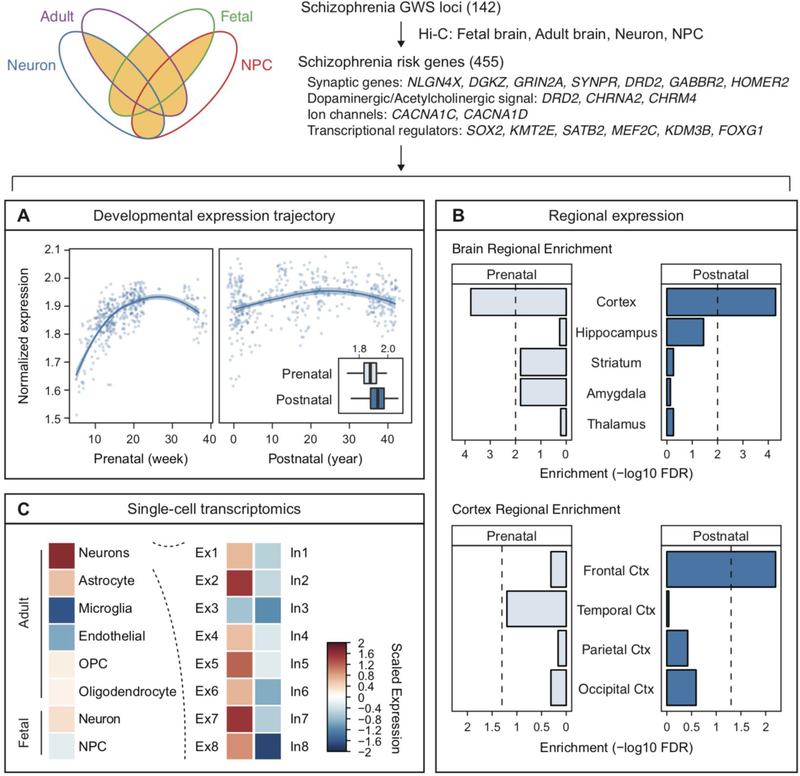
Figure 4. Cellulo-spatio-temporal resolution of Hi-C defined schizophrenia risk genes.
Schizophrenia risk genes were defined by having more than two sources of Hi-C evidence from adult brains, fetal brains, neural progenitor cells (NPC), and neurons. A. Developmental expression trajectory of Hi-C defined schizophrenia risk genes suggests midgestation and postnatal periods as critical windows. B. Regional enrichment of schizophrenia risk genes in the frontal cortex. C. Single-cell expression values of schizophrenia risk genes highlight excitatory neurons as central cell types. Ctx, cortex; Ex, excitatory neurons; In, inhibitory neurons; OPC, oligodendrocyte precursor cells.

