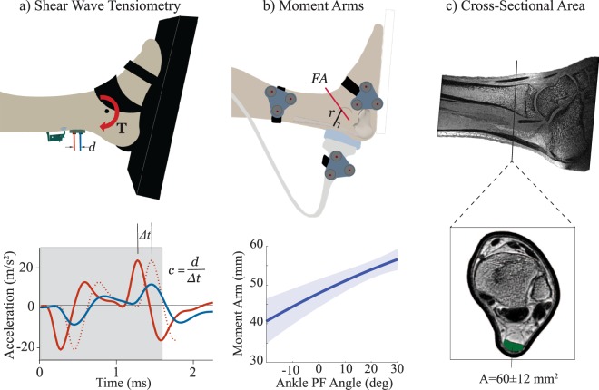
Figure 1.
(a) Subjects performed cyclic isometric exertions while two accelerometers measured the skin motion associated with an induced shear wave propagating in the tendon. Cross-correlation of the signals within an adaptive window (see gray box that includes acceleration peaks induced by the tap event) was used to determine the propagation time Δt, and hence wave speed c. (b) Coupled ultrasound and motion analysis collections were used to characterize the Achilles tendon moment arm, r, as a function of ankle plantarflexion (PF) rotation about a functional axis (FA)13. (c) MR images were segmented to compute the Achilles tendon cross-sectional area, A, at the location where the accelerometer array was placed.

