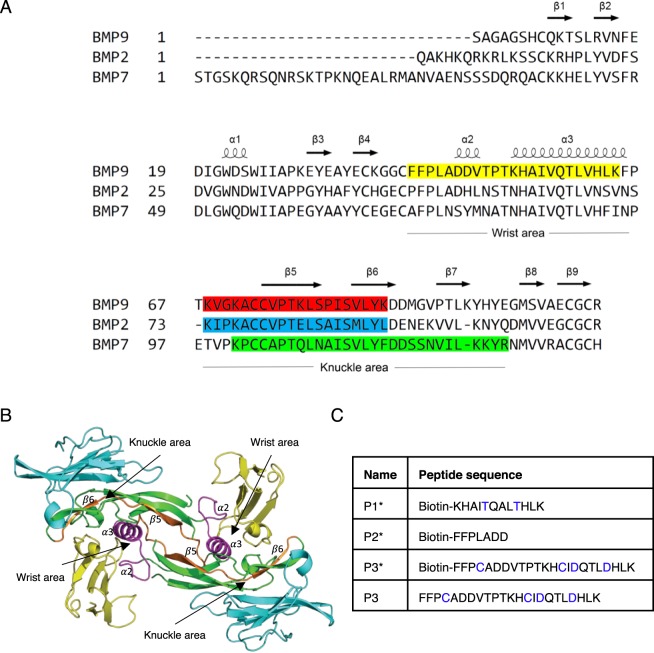
Figure 1.
Design of BMP peptides. (A) Sequence alignment of BMP9, BMP2 and BMP7 with the secondary structures, “knuckle area” and “wrist area” annotated. Previously reported BMP peptides are highlighted in red, blue and green. P3 sequence in the current study is highlighted in yellow. (B) The crystal structure of ALK1:BMP9:ActRIIb (4FAO)8. BMP9 in green, ALK1 in yellow, ActRIIb in cyan. The P3 peptide, which is designed from the wrist area of the ALK1-binding surface, is highlighted in magenta. The P4 peptide (Fig. 3), which stretches across the knuckle surface of the BMP9 is highlighted in orange. (C) The peptide sequences of P1*, P2*, P3* and P3.

