Figure 1.
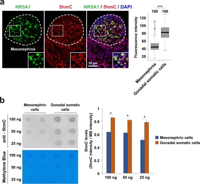
5-hydroxymethylcytosine is preferentially enriched in NR5A1-positive gonadal somatic cells. (a) Co-immunostaining profiles of NR5A1 and 5hmC in the central regions of XY E11.5 gonads. Enlarged boxes indicate co-localization of NR5A1 and 5hmC in gonadal somatic cells. Fluorescence intensity values of 5hmC in every 100 gonadal somatic cells and mesonephric cells were examined and summarized in a box plot (right). Signal intensity was quantified using ImageJ software. ***P < 0.001. n = 100. (b) Comparison of 5hmC amounts between mesonephric cells and gonadal somatic cells. Mesonephric cells and gonadal somatic cells were separated from XY embryos at E11.5 that carried Nr5a1-hCD271-transgene (see Materials and Method section) and were introduced into Dot-blot analysis. The relative amounts of 5hmC were calculated by dividing the signal intensity of 5hmC by that of methylene blue (right). Signal intensity was quantified using ImageJ software. Error bars indicate the SEM values of duplicates of the dot signal intensity. *P < 0.05.

